
Introduction
Despite its at times challenging nature, electrocardiography is an extremely useful diagnostic modality in the evaluation of cardiovascular disease. Electrocardiography is specifically most valuable for evaluation of patient heart rate and rhythm, however also allows gives information regarding cardiac chamber enlargement, pericardial disease, and various metabolic conditions such as hypoxia and electrolyte abnormalities. Proper understanding of the diagnostic utility of ECG requires basic knowledge of the normal cardiac electrical system.
Normal Cardiac Conduction
Myocardial activation via the normal cardiac conduction system results in sequential activation of the atria and ventricles, allowing for an organized and efficient cardiac contraction. This allows for proper filling and emptying of the heart to ensure appropriate cardiac output. The conduction system of the heart is composed of:
- Sinoatrial (SA) node
- Atrial internodal fibers
- Atrioventricular (AV) node
- Bundle of His
- Right and Left Bundle Branches
- Purkinje fibers
The cells of the SA node, AV node, and Purkinje fibers all have the properties of automaticity, with the SA node demonstrating the most rapid rate of spontaneous depolarization (i.e., 60-180 bpm in dogs, 160-240 bpm in cats). The wave of depolarization initiated by the SA node is propagated via the internodal tracts through the atrial myocardium (resulting in the P-wave), and ultimately to the AV node.
The AV node is located in the interatrial septum just above the ventricles and is the only normal conduction tissue from the atrial to ventricular myocardium. Due to its small size, depolarization of the AV node cannot be viewed as waveform on the surface ECG. As conduction velocity through this tissue is relatively slow (compared to atrial and ventricular myocardium), the duration of the ‘P-R interval’ on the surface ECG is largely contributed to by AV nodal depolarization. The AV node also normally demonstrates the property of automaticity, and cells in this region are capable of spontaneous depolarization in the event of failure of SA nodal depolarization however this occurs at a slower rate (i.e., 40-60 bpm in dogs, 100-150 bpm in cats).
Following activation through the AV node, the Bundle of His and bundle branches conduct impulses to the Purkinje fibers and ventricular myocardium. Conduction velocity in His, bundle branch, and Purkinje fibers is extremely rapid, and the Purkinje fibers also demonstrate spontaneous automaticity. With failure of SA and/or AV nodal stimulation, the Purkinje fibers can depolarize to result in ventricular contraction albeit at a much slower rate (i.e., 20-40 bpm in dogs, 70-120 bpm in cats). When depolarization of the ventricular myocardium occurs through stimulation following AV nodal, His, and bundle branch activation, a normal QRS complex is visualized on the ECG. With ventricular depolarization via Purkinje fiber automaticity without using the normal cardiac conduction system, a wide and bizarre QRS complex occurs.
ECG Interpretation
Although pattern recognition may allow for the identification of obvious arrhythmias, proper evaluation of the ECG requires a systematic approach to ensure accuracy. The following ECG characteristics should be evaluated with every diagnostic ECG:
- Recognition of ECG settings
- Determination of heart rate
- Evaluate cardiac rhythm
- Measure amplitude and intervals
- Determination of Mean Electrical Axis (MEA)
- Assess for chamber enlargement patterns
Did you know that Oncura Partners has a dedicated cardiology team that can review your ECGs? Click here to request more information about our electrocardiography support services.
Normal Cardiac Rhythms
Sinus Rhythm
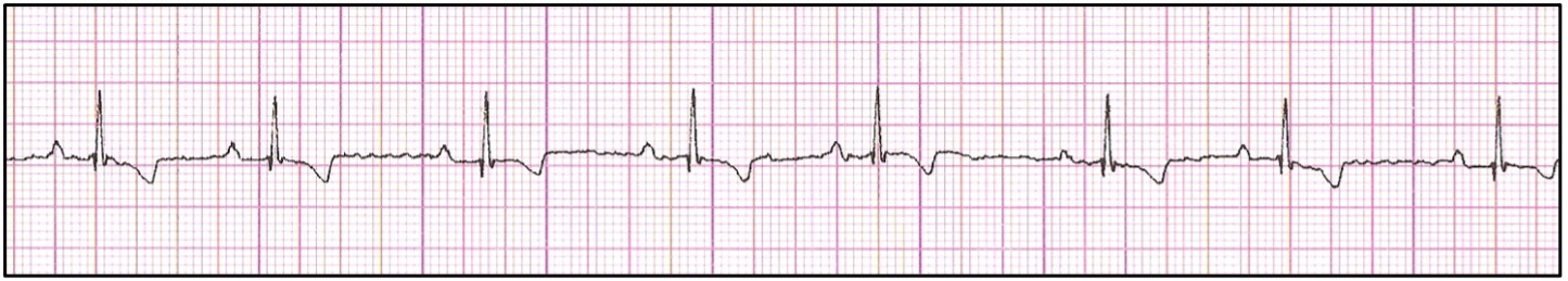
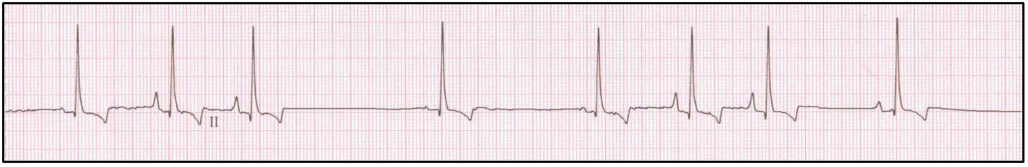
A sinus rhythm represents the normal sequence of cardiac electrical activity. Based upon normal P-QRS-T morphology and relationship, consistent P-P and R-R intervals, and normal heart rate, activation of atrial and ventricular myocardium following initial depolarization of the SA node and propagation through the AV node and His-Purkinje system is assumed. The rate of a sinus rhythm varies between species (i.e., dog – 60-180 bpm, cat 160-240 bpm).
Sinus Arrhythmia (and Wandering Pacemaker)
A sinus arrhythmia results from phasic variation in vagal (parasympathetic) tone. Although this often in conjunction with the respiratory cycle (with increasing heart rate upon inspiration, and decreasing with exhalation), other causes of increased vagal tone are also possible. This ‘arrhythmia’ is considered a variant of normal in dogs however is typically associated with disease entities resulting in increased parasympathetic tone in cats (i.e., gastrointestinal, respiratory, and/or CNS disease).
A sinus arrhythmia is often accompanied by the phenomenon referred to as a ‘wandering pacemaker’ in dogs. Although this title is often inaccurately associated with a pathologic arrhythmia, a wandering pacemaker is the result of depolarization of different locations within the SA node due to changes in vagal tone. This results in slight differences in the vector of depolarization of atrial myocardium, thus affecting the P-wave morphology. Typically, P-wave shape increases with increasing heart rate (within inspiration) and decreases with decreasing heart rate (with exhalation).
Abnormal Cardiac Rhythms
Abnormal cardiac rhythms (bradycardia and tachycardia) will be covered in Part 2 and Part 3 of this blog series on electrocardiography.