
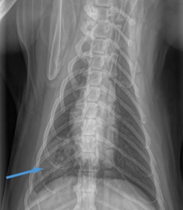
“Sasha” is a 12-year-old F/S DSH who was referred for a 2 week history of inappetance and weight loss. On physical examination, she was thin with an unkempt coat and a II/VI systolic murmur. Initial evaluation included CBC, chemistry profile, UA and thoracic radiographs. Thoracic radiographs showed a normal cardiac silhouette and a focal region of patchy infiltrates in the right caudal lung lobe. A thoracic ultrasound was requested.
Figures 1,2: Thoracic ultrasound with Vet BLUE lung scan revealed a focal shred sign in the right caudal lung lobe consistent with a consolidated/infiltrative lung lobe lesion. In addition, several small (3-5 mm) nodules were noted in the right cranial lung lobe region. Echocardiogram was normal.
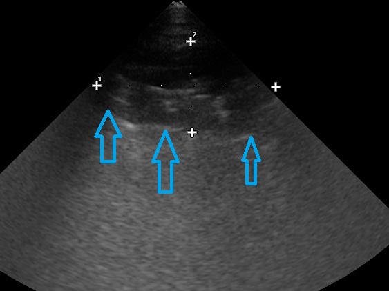
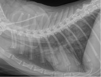
Figure 3: Shred sign right caudal lung lobe region.

Figure 4: Lung nodule (5 mm) detected with Vet BLUE in the right cranial lung.
Vet Blue Lung Scan: A Vet Blue lung scan was performed in the order of caudodorsal lung lobe region, perihilar lung lobe region, middle lung lobe regions, and lastly cranial lung lobe region on the left and right hemithorax over single representative intercostal spaces. BLUE stands for Brief Lung Ultrasound Exam.
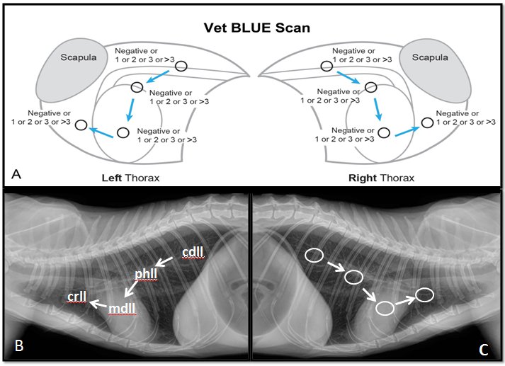
- Left thorax: 0, 0, 0, 0; no evidence of interstitial lung fluid (pulmonary edema) or other lung pathology
- Right thorax: Large shred, 0, 0, multiple small nodules in the right cranial lung lobe region
- Interpretation: There is a large region of focal consolidation in the right caudal lung lobe region (represented by the large shred sign) without obvious evidence of associated pulmonary edema. This lesion measures approximately 2.7 x 1.4 cm and contains entrapped air consistent with lung tissue. There is no evidence of interstitial edema associated with the lesion. There is evidence of multiple focal nodular lesions in the right cranial lung region.
Fine needle aspirates were performed of the right caudal lung lobe mass. Cytology was consistent with pulmonary carcinoma. Subsequently, a CT of the thorax was performed to evaluate for metastasis. In addition to the primary lung lobe mass, several small metastatic nodules were seen in the right cranial lung lobe on CT.
Vet BLUE can be extremely valuable for assessing for lung pathology that is questionable on thoracic radiography. Several small nodules were detected on the initial Vet BLUE exam although they were not evident radiographically. Therefore, the initial assessment of the thoracic ultrasound included “the nodules in the right cranial lung region are suspicious for metastasis.” In addition, in the past, I have been reluctant to perform fine needle aspirates on lung lesions that were not discrete masses on thoracic radiographs. In fact, I did not recommend performing lung ultrasound on lung lesions unless the lesions were discrete masses along the lung periphery. Now that I am routinely incorporating Vet BLUE into my thoracic evaluations, I find that I am able to perform fine needle aspirates on far more lung lesions than I have in my past 20 years of practice. I now realize that I have another valuable method to assess lung pathology that often provides additional information to traditional radiography.
Images from the textbook Focused Ultrasound Techniques for the Small Animal Practitioner, Wiley 2014. Updated Edition 2020: Point of Care Ultrasound Techniques for the Small Animal Practitioner. Editor, GR Lisciandro.