
Diagnosing pancreatitis can be challenging in dogs. Ultrasound assessment can be a useful diagnostic tool when it comes to diagnosing pancreatitis along with clinical signs of acute onset of vomiting, nausea, hyporexia, lethargy, abdominal pain and pancreatic lipase elevation. The pancreas is an elongated, thin, lobular organ with a right limb, left limb and body. The normal appearance of the pancreas is isoechoic to mildly hypoechoic compared to the surrounding mesentery and is less than 1cm in width.
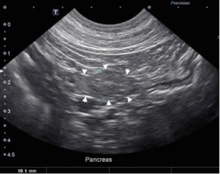
Figure 1: Normal canine pancreas seen between the white arrowheads1.
Pancreatitis can have multiple ultrasound abnormalities and variable appearances. The appearance of acute pancreatitis is an enlarged or thickened, hypoechoic pancreas with peri-pancreatic hyperechoic fat. The pancreas can become rounded with irregular margins and heterogenous echotexture. Dilation of the pancreatic duct, biliary duct, regional peritoneal effusion, GI ileus, gastric thickening and mild corrugation of the duodenal wall can also be seen.
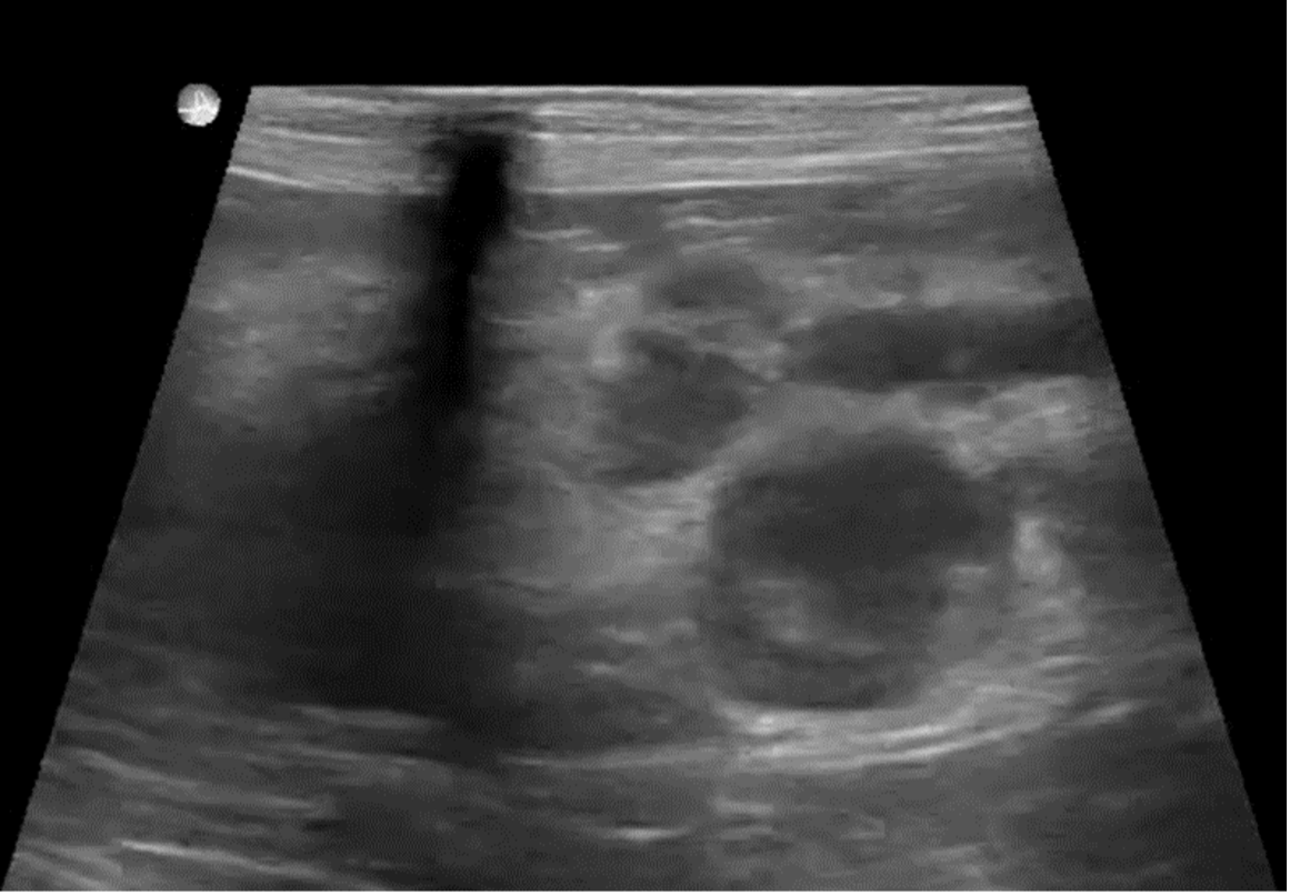
Figure 2: Pancreatitis of the left limb. Rounded, hypoechoic pancreas with mild regional hyperechoic fat.
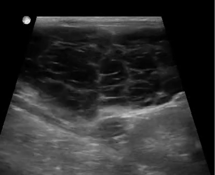
Figure 3: Severe pancreatitis that appears mass like. Enlarged, hypoechoic, irregular, nodular pancreas.
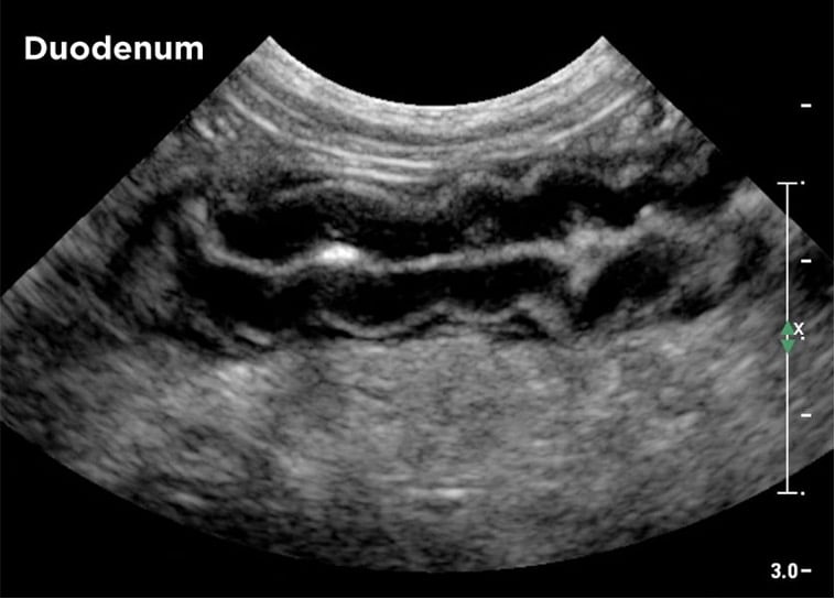
Figure 4: Duodenitis secondary to pancreatitis of the right limb. Corrugation of the duodenum with surrounding hyperechoic mesentery 2.
Dogs with pancreatitis can have one or any combination of these abnormalities and findings may be present in one or both limbs. It is also important to remember that a normal appearing pancreas does not rule out pancreatitis. Ultrasound should not be the only criteria used to diagnose pancreatitis. Ultrasound is also useful in ruling out other potential causes of abnormal clinical signs and lab abnormalities.
References:
- Tsai, S from The Role of Imaging in Diagnosing Pancreatitis accessed 21 June 2022 <://www.mspca.org/wp-content/uploads/2015/11/Image-2-300x237.jpg>
- Huyn, E and Berry C. 2018 May/June, from Today’s Veterinary Practice, Small Animal Abdominal Ultrasound: The Pancreas, accessed 21 June 2022 https://todaysveterinarypractice.com/wp-content/uploads/sites/4/2018/04/T1805C01Fig12-768x551.jpg