
Ultrasound assessment of patients is extremely important in the acute veterinary setting. Point of care ultrasound (Global FAST) should be used in addition to thorough physical exam and laboratory evaluations to offer the timeliest diagnosis and therapeutic intervention in critical patients. Ultrasound assessment of the gallbladder has proven to be an important tool. The finding of gallbladder wall edema (GBWE) in acutely ill patients can be a marker for anaphylaxis, right sided heart failure, pericardial effusion, or acute cholecystitis. Other documented causes include hypoproteinemia, immune-mediated anemia, sepsis, pancreatitis, and post-transfusion (likely immune-mediated; possibly volume overload).
A recent article in the Journal of Veterinary Internal Medicine entitled, “Thirteen dogs and a cat with ultrasonographically detected gallbladder wall edema associated with cardiac disease” (JVIM 2021; Lisciandro, et al) identified the presence of gallbladder wall edema in patients with varying instances of cardiac disease. Knowing this significant ultrasound finding is especially important in the emergent setting since patients with anaphylaxis and cardiac disease may have similar presentations including acute weakness and collapse.
It in this study, gallbladder wall edema is described as an ultrasonographically striated gallbladder with mural thickening ranging from 3-5 mm. This finding is also commonly known as the gallbladder halo sign. Of the dogs, 11 of the 13 had pericardial effusion, one had dilated cardiomyopathy and one had right ventricular myocardial failure. In addition, 9 of the 13 dogs had ascites. One cat was also included in study and this patient had right sided heart failure associated with a ventricular septal defect; the cat also had ascites.
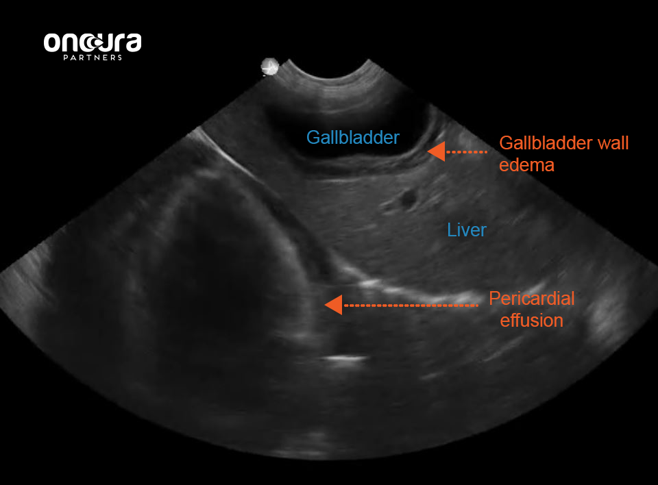
In patients with acute weakness and/or collapse, ultrasonographic assessment of the gallbladder is very important. Patients with ascites are often presented for abdominal ultrasound and thoracic pathology is missed since these animals often do not have heart murmurs. It is important to remember to look past the diaphragm at the subxiphoid site when evaluating the liver to look for pericardial effusion. It is also important to assess these animals for distention of the caudal vena cava and hepatic veins. In animals with a gallbladder halo sign secondary to anaphylaxis, the caudal vena cava is often small or collapsed due to decreased venous return. A distended vena cava with lack of bounce (due to the dynamic changes during cardiac and respiratory cycles), supports the presence of cardiogenic gallbladder wall edema. The point of care protocol for Global FAST (AFAST, TFAST, Vet BLUE) incorporates a standardized assessment of all of these findings to allow a more accurate diagnosis of pericardial effusion or right sided heart failure versus other causes of gallbladder wall edema.
Ultrasound assessment is a valuable tool for assessment, especially in the emergent setting, but I would advise ultrasound assessment in any sick animal as an extension of the physical exam.
Did you know that as an Oncura Partner Clinic, you can submit your ultrasound results along with other diagnostics including radiographs and bloodwork for fast, board-certified specialist interpretation? Click here to request more information about Oncura's unique ultrasound platform and telehealth services.
References:
1. Lisciandro GR, Gambino JM, Lisciandro SC. Thirteen dogs and a cat with ultrasonographically detected gallbladder wall edema associated with cardiac disease. J Vet Intern Med. 2021;35: 1342–1346. https://doi.org/10.1111/jvim.16117
2. Lisciandro GR. Abdominal and thoracic focused assessment with sonography for trauma, triage and monitoring in small animals. J Vet Emerg Crit Care. 2011;21(2):104-122.
3. Nelson NC, Drost WT, Lerche P, et al. Noninvasive estimation of central venous pressure in anesthetized dogs by measurement of hepatic venous blood flow velocity and abdominal venous diameter. Vet Radiol Ultrasound. 2010;51(3):313-323.